Quantitative measurement and imaging of drug-uptake by bacteria with antimicrobial resistance
Short Name: MetVBadBugs, Project Number: 15HLT01
Innovative imaging techniques to help treat drug resistant pathogens
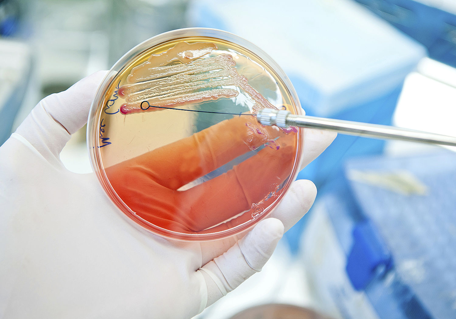
In Europe, antimicrobial resistance is responsible for around 25 000 deaths a year with annual treatment and social costs estimated at around €1.5 billion. Two thirds of these deaths are caused by Gram-negative bacteria, so called as they fail to retain the crystal violet dye used in the Gram staining procedure. These bacteria have a double membrane and ‘protein pumps’ that rapidly remove antimicrobial drugs that penetrate the cell before they can take effect. In addition, these bacteria can also attach to surfaces, such as implanted medical devices, and form dense aggregations called ‘biofilms’ which can be 100 times more resistant to antibiotics than singly growing, or planktonic, cells.
To treat these bacteria requires knowledge on how such biofilms are organised. Analysis is problematic however as the biofilms surrounding bacteria contain high amounts of water meaning that many high-resolution analytical techniques, such as Secondary Ion Mass Spectrometry (SIMS), cannot be utilised as these are vacuum based techniques and require water free samples. However, dehydration led to the 3D structure of such films being compromised meaning that information on how the bacteria are organised and resist treatment was lost.
This project advanced the measurement capability to treat drug resistant bacteria by providing the metrology to quantitatively measure and image the localisation of antibiotics and to understand the penetration and efflux processes in bacteria and biofilms.
A High-pressure freezing procedure was developed to allow analysis of planktonic bacteria and biofilms up to 500 μm thick. This method prevented the formation of ice crystals in samples which can cause disastrous effects on biofilm structure and allowed imaging of biofilms in their near-native state using the cryo-OrbiSIMS instrument. This technique has been published in a Good Practice Guide.
Procedures were also developed for two types of Raman spectroscopy, ‘tip enhanced Raman scattering’ (TERs) and ‘surface-enhanced Raman spectroscopy’ (SERS) which measure the vibrational modes of molecules to provide a structural fingerprint, allowing identification.
The knowledge developed in the EMPIR project has been transferred to the UK’s National Biofilms Innovation Centre which brings together researchers from multiple disciplines in the UK, across academia and industry, to prevent, detect, manage and engineer biofilms.
As well as increasing understanding on how bacteria resist treatment the methods developed in the project are also applicable to viruses like SARS-CoV-2, raising the possibility these techniques might be used for fighting both bacteria and viruses.
Coordinator: Paulina Rakowska
For more information, please contact the EURAMET Management Support Unit:
Phone: +44 20 8943 6666
E-mail: empir.msu@euramet.org
Biomedical Imaging and Sensing Conference
Analytical Chemistry
RSC Advances
AAPS PharmSciTech
RSC Advances
Biophysical Journal
Surface and Interface Analysis
Analytical & Bioanalytical Chemistry
