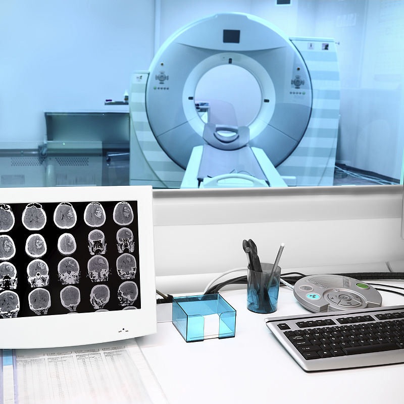
Trusted MR-guided radiotherapy planning
Challenge
Europe’s health services face demands for breakthrough cancer therapies assured as safe, personalised, and able to cost-effectively treat patients.
In 2020, 2.7 million people in the European Union received a cancer diagnosis. About half underwent radiotherapy, including during diagnosis. Image-guided radiation therapies became common for diagnosis and treatment, combining therapy and imaging technologies in one device. Computed tomography emerged as the dominant imaging technology but its use of multiple X-rays limits how much imaging can be used.
By contrast, MR-guided radiotherapy (MRgRT), which combines Magnetic Resonance Imaging (MRI) with high-energy photon or X-ray beams for therapy, offered zero additional radiation exposure and so unlimited imaging. The higher soft-tissue contrast of MRI also provides easier differentiation between healthy and cancer cells, and continuous imaging aids correct positioning during treatment. Combined, these advantages offered better patient outcomes and reduced side effects.
Elekta, a medical technology company and leader in precision radiation therapy, along with University Medical Center Utrecht (UMCU), Phillips and others developed an MRgRT (or MR-linac) concept, installed as the world’s first clinical prototype in 2014. The Elekta Unity was designed so clinicians could see tumours and surrounding healthy tissues as treatment doses are delivered, so can effectively be adapted for changes in patient anatomy between sessions.
Successful clinical radiotherapy depends on delivering accurate radiation doses to patients to the right area - typically supported by trusted codes of practice. However, magnetic fields, which are part of the working principle of MRI scanners, were shown to interact with treatment beams and measurement equipment. The Elekta Unity was designed to mitigate effects on the generation of the treatment beam, but the effect of magnetic fields on measurement devices used by clinicians for calibrating and characterising radiation fields as part of treatment planning was unknown, and not described in codes of practice.
Elekta developed its treatment planning software (TPS) so clinicians could calculate optimal dose distributions for each treatment session, the accuracy of which was relied on an underlying beam model and measured dose input data. While prototypes of the Unity showed the capability of accurate dose delivery, for Elekta to be confident that personalised doses could be delivered in practice, strong evidence was needed to show the reliability of its TPS.
Solution
The EMPIR project Metrology for MR guided radiotherapy developed dosimetry and imaging metrology supporting safe clinical implementation and innovation in MRgRT.
Changes in characteristics of radiation detectors and radiation fields with magnetic field strength were investigated through measurement and simulation. A film-based approach was developed replacing water tank measurements for dose input data measurement for MRgRT treatment planning systems. Data was fed into Elekta’s TPS, and a beam model devised, providing measurements usable for commissioning Elekta Unity devices.
Correction factors for detectors used to calibrate reference fields in MRgRT facilities were established by measurement and simulation. Quality assurance procedures for MRgRT facilities were also developed for static and dynamic conditions, including tests of clinical workflows with intra-fraction motion based on motion phantoms, and methods to track organ motion during treatment.
Impact
The new beam model was used in the first trial treatments with the Elekta Unity in 2017. Supported by the Dutch Cancer Society, this First in Man study showed the clinical feasibility of MRI guided radiotherapy. Verification of the accuracy of algorithms in its TPS showed that prescribed doses could be accurately delivered to patients in clinics, considered an essential prerequisite for commercial success.
Approval for European commercial sales and clinical use was granted in June 2018. Two months later Elekta announced a first clinical treatment, of a patient with long term prostate cancer. UMCU used an Elekta Unity to visualise soft tissue targets before and during each treatment, allowing daily adaptation of the treatment plan with changes to the patient’s anatomy. A key benefit was cutting the time to create the treatment plan from
days to a few minutes.
Providing trust in treatment planning advanced the development and clinical acceptance of MRgRT, heralding more precise and personalised treatments, shorter treatment times and reduced cost of care for healthcare providers.
- Category
- EMPIR,
- Health,
