Spotting inter-cell communications
Challenge
Many diseases, including cancers, diabetes and heart disease increase the number of small cell particles, extracellular vesicles (EV) or microvesicles in the bloodstream. These have recently been discovered to both indicate disease and also play an important role in spreading cancer around the body. Being able to make earlier disease diagnosis, monitor the effectiveness of treatments, and potentially deliver drugs to target cells could all be possible by measuring and using blood borne EV.
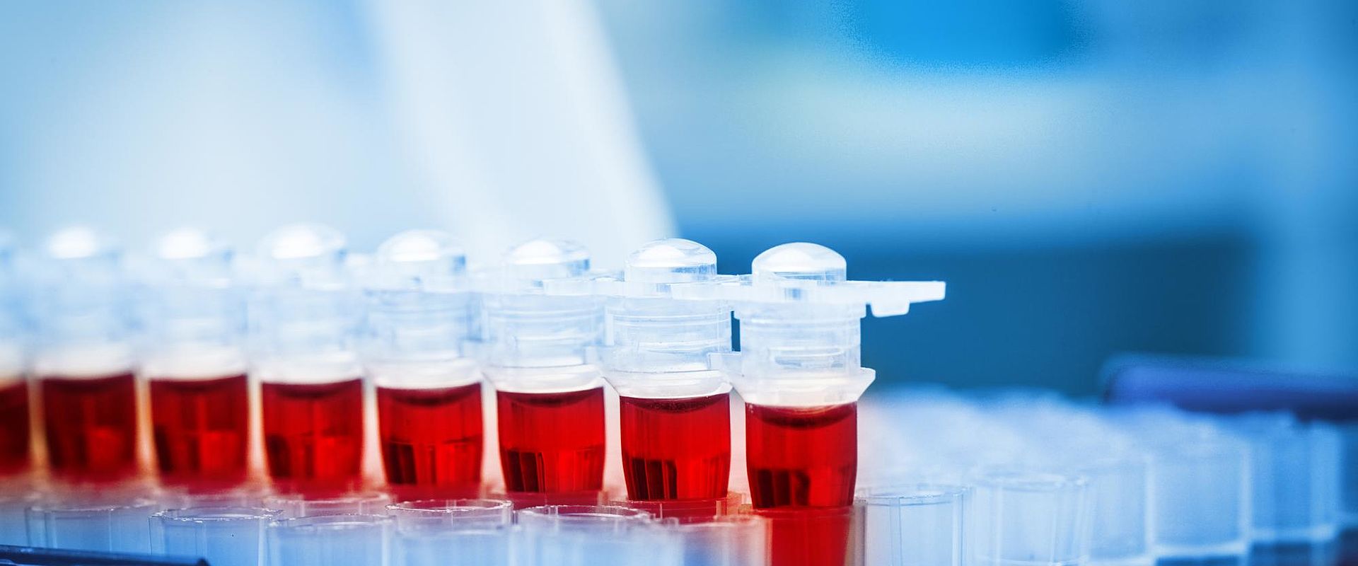
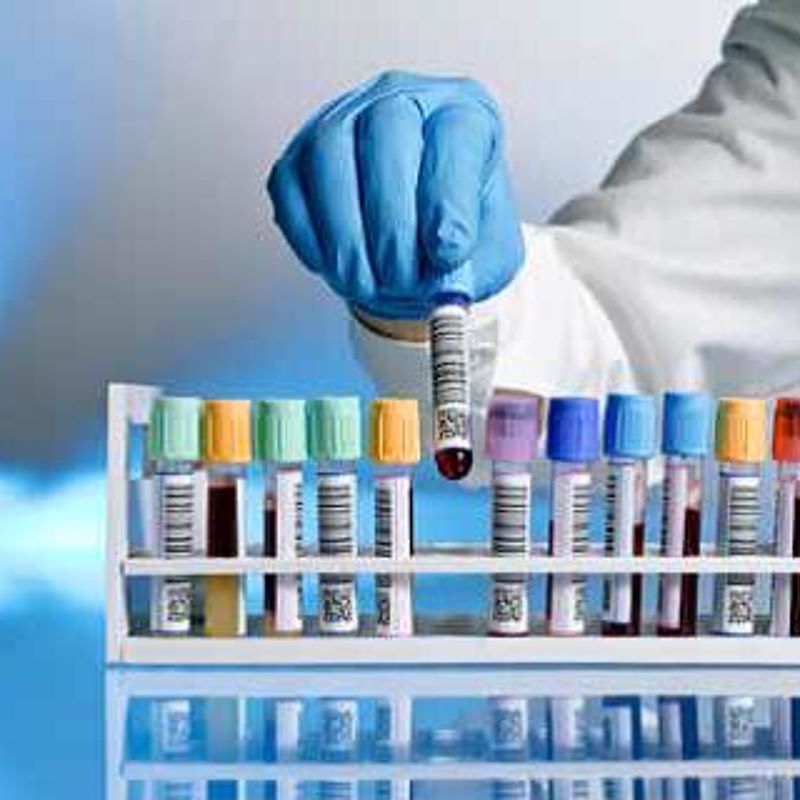
Before EV can be used for any of these purposes, we need reliable, reproducible and standardisable ways to measure them. This presents a number of challenges, the first of which is preparing high quality blood samples for measurement. Preparation methods must ensure that EV are retained in the sample for analysis whilst contaminating molecules that will interfere with EV detection are removed. Reliable, accurate, standardised methods are needed for collecting samples, separating out the small EV particles, and storing them before measurement.
Solution
The EMRP Project Metrological characterisation of microvesicles from body fluids as non-invasive diagnostic biomarkers identified optimal procedures for EV blood sample collection, and its preparation and storage prior to analysis. The project developed a recommended approach for EV collection and purification. It identified that rapid freezing in liquid nitrogen and maintaining at -80 °C was important in ensuring samples did not deteriorate before analysis.
One of the most important findings of the project was that traditional separation methods, which involve ultra-centrifugation - spinning samples at very high speed – has problems that prevent the production of reproducible samples. The team found that ultra-centrifugation leads to the loss of some EV particles; extreme pressures experienced in repeated spin cycles can damage EV particle structures and collection of other materials along with EV can affect result accuracy.
The project demonstrated that size exclusion chromatography (SEC) - a separation method in which the sample is filtered through a ‘mesh’ – is a robust and reproducible EV sample separation method, which produces pure EV samples. This has the potential to open up the use of EV for disease diagnosis.
Impact
Izon Science, a manufacturer of precision instrumentation for nano- and micro-scale particle analysis, has taken the project’s SEC EV separation method and generated an easy to use kit for clinics and medical researchers. The kit is inexpensive and only requires 15 minutes to produce a pure EV sample, a huge advance on the 2-3 hours required by the less accurate centrifuging technique.
Over 250 labs worldwide are now using the improved EV sample preparation methods developed in the project - highlighting the growing interest in these micro-particles for medical research. The project’s sample preparation methods have been incorporated into good practice guidance being drafted by the leading international bodies involved in EV research. This is a first step towards a new ISO standard for EV separation and analysis which will lay the foundations for using EV measurements in new point of care diagnostic devices.
EV research, especially that examining communication between cells, is advancing rapidly, and holds huge promise in the diagnosis and treatment of many serious diseases. The approaches identified by this project, and the kits commercialised by Izon, will be critical to ensuring the quality of samples on which this research depends.
- Category
- Health,
- EMRP,
