New reference material for light scatter-based sizing of extracellular vesicles
Challenge
Extracellular vesicles (EVs) are biological nanoparticles actively released by cells, comprising of an outer lipid membrane, and containing markers indicative of the cell of origin, and housing an inner aqueous core. Most EVs are 100 nm in diameter or less but can range in size from 30 nm to a few micrometres. EVs are found in all body fluids. Moreover, cells affected by disease release altered EV profiles compared to normal cells. Therefore, there is a great clinical interest in the use of EVs as biomarkers to diagnose a wide range of medical conditions.
A common technique used in hospitals to characterise EVs is flow cytometry (FCM). FCM can analyse thousands of individual EVs per second by measuring the light scattered as EVs pass through a laser beam, providing data on size, concentration, granularity, and fluorescent intensity.
However, an EV sample characterised on one cytometer is unlikely to return the same results on a different cytometer. As well as different optical configurations, flow cytometers are commonly calibrated for EV measurements using polystyrene beads with a solid core and a high refractive index (RI, ~1.63) compared with EVs (≤ 1.40). As a result, polystyrene beads scatter light 1000-fold more than similar-sized EVs, leading to misinterpretation of light scattering signals of EVs and making comparisons of clinical results difficult.
Solution
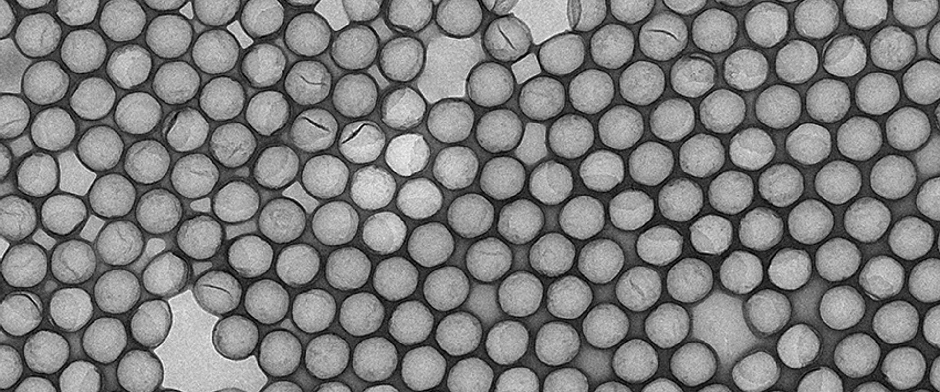
During the METVES project, a new reference material for EVs was synthesised by combining a basic amino acid catalysis route with a hard template approach. Silica beads of 200 nm and 400 nm were coated with 1,2-Bis(triethoxysilyl)ethane to form an organosilica ‘shell’ over the particles. The inner silica core was then ‘etched away’ to generate Hollow Organosilica Beads (HOBs).
Flow cytometry two-angle light-scattering measurements then confirmed HOBs had light-scattering properties and an effective RI similar to platelet-derived EVs (≤ 1.40). However, during the project, HOB preparations were small scale and problems were encountered in reproducibility of the organosilica shell thickness (5-10 nm).
The METVES II project developed procedures to scale up HOB production ten-fold. ‘Small-angle X-ray Scattering’, a nano-scale measurement technique, was used to obtain the diameter and shell thickness of HOBs. This demonstrated the reproducibility of the core size and the shell thickness (~ 10 nm). Nanoparticle tracking analysis and ‘single particle inductively coupled plasma mass spectrometry’ confirmed the HOBs concentrations. A protocol was also established to label the HOBs with the fluorescent compound fluorescein isothiocyanate, a commonly used fluorochrome to label EVs.
Impact
Founded in 2015, the goal of Exometry BV, based in the Netherlands, is to promote the use of EVs as biomarkers of disease by applying physics to both understand and push the technological limits involved in EV detection.
The company was a project partner in METVES II, which allowed them access to the technology and metrological knowledge to generate traceable data on the HOBs they were developing. After the end of the project in 2022, the company commercialised this new reference material as “Verity Shells”, which are applicable for use on all flow cytometers with a scatter detector capable of detecting at least 100 nm polystyrene beads.
The development of more realistic calibration material, such as the Verity Shells, will allow the validation and reproducibility of EV measurements from clinical samples. In turn, this approach will allow cheap and rapid diagnostic tests to replace the more expensive and invasive tests such as solid biopsies and also allow
earlier identification of pathological conditions in patients.
