Improving radiotherapy success
Challenge
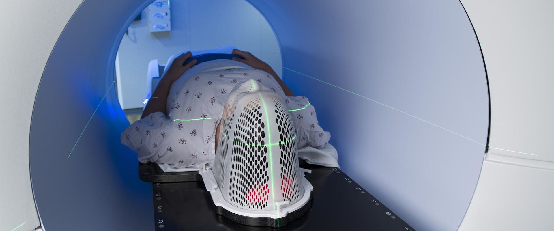
Radiotherapy has been a mainstay of cancer treatment for over a century. It most commonly involves using a linear accelerator (linac) to deliver high-energy beams of X-ray radiation to patients, killing cancerous cells by damaging their DNA. Prior to treatment, patients are imaged in a CT scanner (using X-rays) to identify the target site. However, the position, size and shape of tumours around the chest and abdomen can change significantly during treatment because of the patient’s breathing, limiting the accuracy with which these tumours can be targeted.
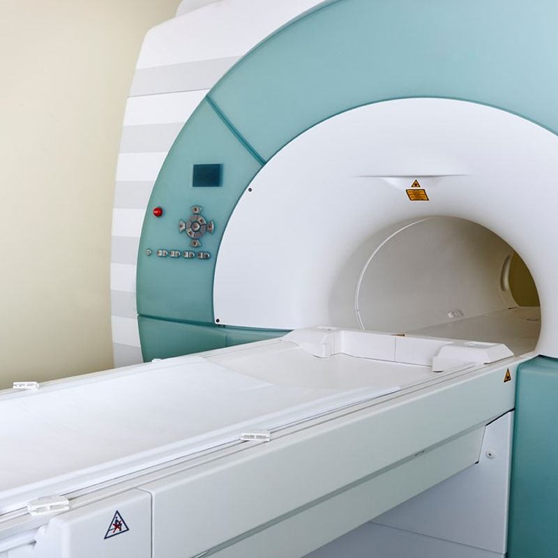
Compared to CT, magnetic resonance imaging (MRI) offers a greater depth of contrast and better visualisation of tissue boundaries, without the ionising radiation. This gives it the potential to provide more detailed images of patients during their treatment. MRI would enable clinicians to track changes to the target site in real time and ensure they are focusing radiation beams as closely as possible on the tumour, improving treatment outcomes for patients.
Delivery of a precise dose of radiation is essential to maximising the success of any radiotherapy treatment, while minimising adverse side effects due to radiation exposure. The problem is the electromagnetic field induced by MRI has the potential to affect a linac’s radiation beam and calibration procedures, and consequently the dose delivered to patients. These effects must be well understood and new methods for calibrating specific MRIlinac combinations developed. This will support the introduction of MRI-guided radiotherapy into hospitals and clinics and improve cancer treatments delivered by external beam radiotherapy.
Solution
The EMRP project Metrology for next-generation safety standards and equipment in MRI developed the first calibration procedure for clinical MRI-guided radiotherapy machines that works in the presence of a magnetic field and allows users to accurately determine the radiation dose delivered to patients. Using a new, compact water calorimeter, MRI-linacs can be calibrated at the hospital and the measurements they make directly linked to national standards. This is a significant improvement over the current calibration method for conventional linacs, in which the linac ion chamber must be calibrated against another ion chamber, itself calibrated at a National Measurement Institute.
Impact
Elekta and Philips, two leading European companies in the fields of radiotherapy and MRI imaging, are part of a consortium developing a combined MRI-linac, which hopes to introduce the new treatment to clinical practice in 2017. The new calibration method developed has enabled Elekta and Philips to calibrate the beam strength and improve beam control of the MRI-linac, enabling customers to have confidence in its ability to provide the right quantity of radiation to a small targeted area. This will support improved treatment of tumour cells, while minimising exposure of surrounding healthy tissue.
The robust and easy-to-perform calibration method developed by the project provides essential support to the safe, effective introduction of the improved image quality offered by MRI into standard radiotherapy treatments. Consequently, the project has made a significant contribution to the development of an innovative, high-value medical technology and the benefits it brings to Europe’s economy and quality of life for citizens.
Speaking of the MRI-linac’s development, Kevin Brown, Global Vice-President of Scientific Research at Elekta, said: “The EMRP project contributes exact and reliable radiation dosimetry to this endeavour, an indispensable precondition before any patient can be treated.”
- Category
- EMRP,
- Health,
