An improved method for studying nanometre-sized biological particles
Challenge
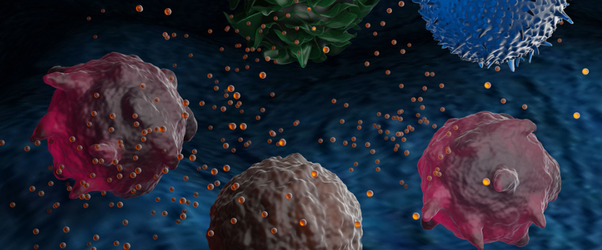
Extracellular vesicles (EVs) are small membrane-coated particles (30 nm - 1,000 nm) released from animal and plant cells. Originally thought to be unwanted cellular debris it has since been realised these are actively secreted, contain soluble molecules such as proteins and nucleic acids, and are involved in both normal and pathological conditions.
Cancerous or virus infected cells often display altered EV morphologies, such as size, refractive index or cargo. EVs can modulate the immune system, cross biological barriers and be engineered to carry specific cargos. These properties make them of great interest in diagnostics, drug-delivery and gene-therapy applications. EV populations are extremely heterogeneous however, and for effective use an accurate characterisation of the subpopulations present is essential.
Many conventional visualisation techniques such as electron microscopy or labelling with fluorescent markers can cause EVs to deform or alter function. Other methods, including nano-flow cytometry or Nanoparticle Tracking Analysis (NTA), cannot image individual EVs over extended times, lack resolving power at the lower size level, or are unable to separate out genuine EVs from debris and other contamination often present in biological fluids.
New techniques were needed to better characterise these tiny particles and unlock their full, medicinal potential.
Solution
During the EMRP project BioSurf, Chalmers University of Technology developed a novel waveguide chip that can be used with standard optical microscopes to detect and study nanoparticles on glass-like surfaces without the usual requirement to label samples with fluorophores. The basic structure of the chip consists of a glass-like core layer embedded in a cladding layer of CYTOP, a polymer material with a refractive index closely matching water (1.34). A small opening is formed in the top CYTOP cladding layer into which a drop containing a nanoparticle sample is placed, directly exposing the particles to the core layer of the waveguide and facilitating interaction with the evanescent part of the light confined within the waveguide structure. The low refractive index of the cladding polymer reduces background light, enabling surface-bound nanoparticles to be discretely imaged with a high signal to background ratio and allowing for label-free detection of faint objects, such as individual sub-100 nm EVs.
Impact
Several years after the end of the BioSurf project in 2015 consortium members founded a new company to commercialise the new waveguide concept – Nanolyze.
Nanoparticles can be analysed in a similar manner to NTA – giving data on EV heterogeneity, size distribution and refractive index at the resolution of single particles. The particles can also be anchored to the surface of the chip giving in-focus analysis periods for single, hundreds, or thousands of EVs over the course of minutes as opposed to seconds for many standard techniques.
Nanolyze further improved the waveguide with the addition of a holding module, a flow system and a fluorescent capability. New, user-friendly software was also designed allowing the easy recording and extraction of data – such as EV uptake into different cell types, interaction with viruses, or the kinetics of drug or RNA loading into the particles.
The company acknowledge that the development of the novel instrument would not have been possible without the BioSurf project and the knowledge of the experts from the measurement institutes involved.
The waveguide will complement existing techniques for nano-sized measurements and give a greater insight into the mechanisms of EVs, helping to unlock their potential for the diagnosis or treatment of numerous pathological conditions.
- Category
- EMRP,
- Health,
- EMN Traceability in Laboratory Medicine,
